
| A Cybernetic Model of Accommodation
Otis S. Brown, McDonnell Douglas Aerospace
Ronald J. Hooker, George Washington University
Peter R. Greene, Harvard University & BGKT Ltd.
Jason S. Moore, The United States Naval Academy
Stirling A. Colgate, The Los Alamos National Laboratory
|
This Paper was delivered to the 1996 Annual Engineering in Medicine
& Biology Society Symposium.
ABSTRACT
Over the past fifteen years we have evaluated numerous models
of accommodation. Our task is to clarify these models by
designing an automatically focused camera, with major emphasis of
the capability of the retina to sense blur and feed this
information back to the eye's lens for accurate focal adjustment.
Depth-of-field, or dead-band, poses a significant obstacle
for the designer of an automatically focused camera. Our approach
is to use noise to provide a scanning, or dither motion so that
the lens will spend 80 percent of its time in sharp focus. Retina
detection of blur can be simulated by a Charge Coupled Device
(CCD), designed to produce a null when sharpest focus is achieved.
The nature of blank-field accommodation is judged, and a
prediction made about its long-term behavior.
INTRODUCTION
This paper's objective is to clarify the predictions that are
implied in earlier block diagrams of the accommodation system.
The diagrams do not provide active outputs which can be compared
directly with the experimental data. The actual building of a
working model from a block-diagram concept is challenging and will
define, after review, the behavior of the normal system.
Thus, for instance, the noise that is seen in the system is
not a defect, but rather is an essential design requirement of a
system that has dead-band. A scanning signal must be present if
the system is to maintain accurate focal control. Other
capabilities of this system, such as blank-field accommodation are
part of the design, and are included in this model. The available
measurements confirm most of the analog computer's predictions for
the eye's dynamic focal control.
THE EYE AS A FOCAL CONTROL SYSTEM
Light rays from objects travel through the cornea, lens and
ultimately arrive at the retina. At the retina they form a
blur-circle which varies in size. The lens control system must
act to drive the lens-plant towards the null (or in-focus)
condition.
THE ORIGIN OF DEAD-BAND, OR DEPTH-OF-FOCUS
Optically, all eyes have dead-band. Dead-band varies, and is
inversely proportional to the size of the aperture. The eye's
dead-band in day-light is approximately +/- 0.6 diopters, and at
night, +/- 0.3 diopters. Dead-band occurs because the lens of the
eye can be varied in power without any detectable change in the
sharpness of the image at the surface of the retina. [1]
A source point of light will produce a blur-circle on the
retina. When this circle is larger than the retina cones, several
cones "fire" producing multiple outputs. As the blur circle is
focused to a point of light, only one cone will fire, producing a
null. This null will exist throughout the dead-band. Because of
this physical characteristic of the eye, a design-around of the
control-system must be accomplished to deal with it, if the system
is to maintain continuous sharp focus on the retina. Dead-band is
schematically shown in Figure 1 and 2.
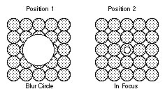
Figure 1: Shows how an expanding "disk of light" or blur circle
falls on a wider area of neurons when the lens is moved
towards and away from the retina. When the disk is
smaller than the diameter of a neuron, only one neuron
is triggered. This depth-of-field is approximately +/-
0.6 diopters.
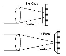
Figure 2: Demonstrates how lens motion relative to the retina
creates variously a "disk-of-light", or blur circle.
For a certain range of motion, no blur is produced on
the surface of the retina.
A FOCUS NETWORK
The basic concept of a null-seeking network and system is
that a cone/neuron will turn-on when struck by a photon of light.
The output of the network depends only on the presence of light or
no-light, and not on its intensity. A Charge Coupled Device (CCD)
could be developed as an analog of this retina characteristic.
[2] (A comparator amplifier, following the cone can be used to
accomplish this conversion of an analog signal to a on-off level.)
Figure 3.
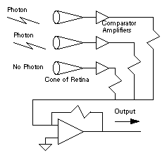
Figure 3: Demonstrates how an "artificial retina" can be designed
to sense blur. While the retina probably uses a more
sophisticated method of determining blur, an auto-focus
camera could be designed and would work using this
basic blur detection strategy.
THE SUMMATION OF NEURONS
When the signal blooms from being out-of-focus (positive or
negative), more cones will detect photons, and the summations of
neuron firing increases. This action produces an increase in
output voltage, as the image goes out-of-focus. Figure 4. Lens
control can occur only after the blur-circle strikes additional
cones.
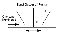
Figure 4: This figure demonstrates the nature of the signal
produced by the artificial retina. Through the area of
dead-band, the signal is constant. When the
blur-circle begins to exceed the edge of the dead-band,
a rising signal is produced.
THE OUTPUT OF THE OPERATIONAL AMPLIFIER
The retina senses the increasing voltage which is used by the
control system (in combination with a dither signal) to produce a
null-seeking action. The dither, or noise will -- on the average
-- center the lens in the middle of the dead-band. This system
will produce sharp focus for perhaps 80 percent of this time when
the eye is viewing an object. Obviously, we are not objectively
aware of the short excursions that occur when the lens exceeds the
edge of the dead-band as the system scans the range of sharpest
focus. Figure 5.
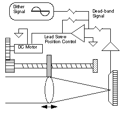
Figure 5: This diagram shows how the accommodation system behaves
in actual operation. Since blur cannot be detected
when the lens is inside the dead-band the focal state
of the lens will "drift" until it exceed the edge of
the dead-band. The system must use negative feedback
to "kick" the lens towards and into the dead-band.
This type of motion is seen with an infrared optometer.
CONSTANT DITHER IN THE LENS SIGNAL
To center the lens, continual lens motion must be induced in
the system. This can be accomplished by a sine-sweep, dither, or
some other noise-type of signal. In the case of the eye, random
motion (noise - from 0.25 to 4 hertz) is seen in lens motion -- as
measured by an infrared optometer. [3,4] The need for this type
of signal should be obvious to most control-system designers,
where static friction or dead-band exist in the control system.
IMPLEMENTATION OF A RETINA-LENS AUTOMATIC CONTROL SYSTEM
The full implementation of the control system requires some
mechanical changes in the "plant" to simulate the eye's behavior.
The manner in which the muscles support the eye's lens has been
discussed in many other texts and dissertations. [5]
Because of a need for high focal accuracy we cannot build a
sphincter-muscle-lens system. We can, however, build a "plant"
that will accomplish the same result. For this model, we show a
lead-screw that is driven by a Direct Current (DC) motor. Thus by
high-gain amplification of the output of the retina, and by
lead-screw adjustment of the lens, we can insure that the lens is
always servoed to the output of the retina for continuous focal
adjustment. Figure 6.
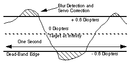
Figure 6: This model of an auto-focus camera shows how the signal
derived from the surface of the retina is used to
control the positioning of the lens relative to the
retina. While more difficult to design, the lens power
could be changed by using the lead-screw to change the
power of the lens -- rather than the position of the
lens. The control-system would behave in the same
manner in either case.
The lead-screw will constantly change the focal position of
the lens and the sharpness of focus on the retina. This type of
signal is seen in the human eye, and is a normal condition. The
continual motion of the lens will cause the retina to provide a
changing signal. Proper use of this signal insures that the lens
is within the dead- band most of the time. The net result of this
control-action is a signal almost identical to the neurological
signal seen when the output of an infrared optometer is recorded
on a strip-chart recorder. [4] A graphic sketch is shown in
Figure 5.
A BLOCK DIAGRAM OF THE SYSTEM
The complete block diagram of the retina-lens system that
captures the basic accommodation characteristic has been
previously published. See Figure 7 [6] The action of the model is
such that when the eye is looking at infinity (zero diopters)
visual environment, the lens will oscillate between +/- 0.6
diopters, as long at the individual looks at infinity.
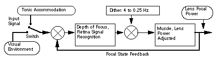
Figure 7: This diagram shows the basic building signal processing
blocks of the accommodation system. The model produces
an output (lens power) that is almost identical to the
measurements made with an infrared optometer.
When the object is moved closer, say to -3.0 diopters, the
lens of the eye will change by +3.0 and then oscillate by +/- 0.6
diopters. Thus we have designed a visual control system that
continuously monitors and tracks its visual environment. This
model is consistent with other proposed dynamic models for the
eye.
ANALOG COMPUTER IMPLEMENTATION OF THE BLOCK DIAGRAM
Our next step is to convert the block diagram into an analog
computer and to compare the output with the response as seen by an
infrared optometer. A major feature of the model is the
requirement that the system must have a stand-by, or reference
position when blur cannot be detected. A control system will
typically drift into the stops, unless a reference signal is
supplied. This simulation is accomplished by a switch which
selects a -1.0 volt level when the eye is in "conscious darkness",
or if blur cannot be detected. A typical value for the dark focus
of the normal eye is -1.0 Diopters. [3] Figure 8.
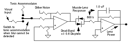
Figure 8: This diagram shows the implementation of the various
accommodation blocks into a design that will work to
effectively control the lens power of the eye.
FUTURE ENHANCEMENTS
The above presentation is a simplified version of
accommodation. In future model enhancements, we will incorporate
a tonic accommodation amplifier which will show that tonic
accommodation will track the average value of accommodation. This
response has been suggested in a previous study. [3] We can also
expect that the tonic accommodation system will show a
time-constant response of approximately 100 days. [7]
CONCLUSIONS
Previous models of accommodation have restricted their
attention to the muscles that surround the lens of the eye. This
model concentrates on the image processing and feedback control
that must occur at the surface of the retina.
ACKNOWLEDGMENT
We acknowledge the long-term assistance of Dr. Karel Montor,
The United States Naval Academy, and Dr. David Guyton, Professor
of Ophthalmology, The Wilmer Institute. Their commitment to the
development of new concepts, and their concern for the welfare of
others, has been an inspiration to all of us. The development of
this paper would have been impossible without their dedicated
support.
CYBERNETICS: [From Gr, kybernetes, steersman, governor.] Comparative
study of the control system formed by the nervous system and
brain and mechanical-electrical communication systems, such
as computing machines.
REFERENCES
1. Hung, G., Ciuffreda, K., Semmlow, J., Hokoda, S., "Model of
Static Accommodative Behavior in Human Amblyopia", Trans. on
Biomedical Eng., Vol. BME-30, No. 10, pp. 665-672, Oct 1984
2. Langenbacher, H. T., Fossum, E. R., and Kemeny, S., "CMOS
Active-Pixel Image Sensor With Intensity-Driven Readout", Jet
Propulsion Laboratory, NASA Tech Briefs, January 1996
3. Baker, R., Brown, B., Garner, L., "Time Course and Variability
of Dark Focus", Investigative Oph. & Visual Science, Vol.
24, pp. 1528 - 1531, Nov 1983
4. Suzumura, A., "Accommodation in Myopia", Department of Oph.,
Aichi Medical U., Proceedings of the 2nd International Myopia
Conference, 1978
5. Greene, P. R., "Mechanical Aspects of Myopia", Ph.D.
Dissertation, Harvard University, Division of Engineering and
Applied Physics, Feb. 1978
6. Semmlow, J., Hung, G., "A Quantitative Theory of Control
Sharing Between Accommodative and Vergence Controllers", IEEE
Transactions on Biomedical Engineering, Vol BME-29, No 5, pp.
364-370, May 1982
7. Brown, O., Young, F., "The Response of a Servo Controlled Eye
to a Confined Visual Environment", The 18th Annual Rocky
Mountain Bioengineering Symposium, pp. 41-44, 1981
Back to  home page...